I heard that cataract surgery is really safe now. Does this mean that cataract surgery no longer has complications?
The advances in technology and surgical techniques has resulted in the high success rates of cataract surgery worldwide. So many people now have had successful cataract operations, and some are now talking about cataract surgery as being only a ‘minor’ procedure.
Does this mean that cataract surgery complications are a thing of the past?
Unfortunately no, complications can still occur during cataract surgery, even in the most experienced of surgical hands.
It is common to have some discomfort, grittiness, sensation of something in the eye, and blood on the surface of the eye after surgery.
The great majority of problems are mild and will clear up over a few days to a few weeks without treatment, or are easily treated without long term consequences.
More serious complications are uncommon and occur in only around 3% of cases. Occasionally, a second operation will be needed to fix problems from the cataract operation.
Severe cataract surgery complications resulting in blindness are very rare (less than 0.1%).
See Related: Patient information guide to cataract surgery
When do cataract surgery complications occur?
Cataract surgery complications can occur during surgery itself, within a few days or weeks of surgery (early complications), and months after surgery (late complications).
Often, these complications occur not because of anything you have or have not done.
If at any point after your surgery, you are concerned that you may have developed any complications, please consult your ophthalmologist without delay. It is better to be safe and get your eye examined. The last thing you should worry about is ‘wasting’ your ophthalmologist’s time.
See Related:
Complications occurring early after cataract surgery
Complications occurring late after cataract surgery
Can you tell me more about the possible complications that might occur during cataract surgery?
All forms of eye surgery carry risk during the actual operation itself, and cataract surgery is no exception.
In this section, I discuss 5 complications that may occur during cataract surgery: hemorrhage, iris trauma, retained lens fragment, posterior capsule rupture, and dropped nucleus.
1. Hemorrhage (bleeding)
There are 3 types of bleeding that can occur during surgery. The most common type is also the least clinically significant. The rarest type is also the most severe clinically.
Subconjunctival hemorrhage: It is relatively common to have a little bleeding or oozing at the front of the eye during surgery. This occurs due to a burst blood vessel in or under the conjunctiva, which can be from the actual operation itself, or from the anesthetic injection prior to surgery.

While subconjunctival hemorrhage from cataract surgery may look serious and unsightly, the blood is located superficially under the conjunctiva and is of no clinical consequence. No treatment is necessary for this. The blood will clear over a week or two.
Hyphema: Sometimes, there may be bleeding into the front chamber of the eye (the anterior chamber). This is usually from a burst blood vessel in the iris or from the ciliary body.
When there is hyphema, your vision will be blurred. The blurring will improve once the blood is cleared from the eye, and this can take a few weeks.
No additional treatment is necessary beyond the usual post-operative eye drops. However, you may need pressure-lowering eye drops if the hyphema has caused your eye pressure to become high.
Suprachoroidal hemorrhage: This type of bleeding is rare (less than 0.1% of the time), but very serious and can cause permanent and irreversible visual loss. This happens when there is a rupture of a large blood vessel between the choroid (middle lining of the eye) and sclera (outer wall of the eye), causing a major bleed behind the eye.
When there is a suspicion of suprachoroidal hemorrhage, the best thing your ophthalmologist can do is to stop the operation immediately and close up the surgical wounds. Continuing the surgery in these circumstances would be exceedingly risky. The operation can be resumed safely after a week or two, once the hemorrhage has settled.
2. Iris trauma
Less than 10% of people have an injury to the iris (colored part of the eye) during surgery. This can happen if the ultrasound probe accidentally sucks in some iris tissue instead of the cataract. This can also happen if pupil enlargement (such as with pupil expanders or iris hooks) was necessary for small pupils that refuse to dilate despite eye drops.
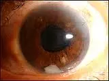
You may notice a discoloration or irregular shape of the iris. Sometimes, you may see stitches inside the eye (occasionally sutures are necessary to repair the iris trauma).
Iris trauma is usually not clinically significant and does not require any active treatment. Sometimes too much light can enter through the damaged iris, causing some visual disturbance.

Iris trauma during cataract surgery may result in an irregularly shaped pupil. It can look quite alarming at first glance. Although generally harmless, it may cause mild symptoms such as glare in bright sunlight.
3. Retained lens fragment in the anterior chamber
During surgery, the cataract is broken up into multiple small pieces. All the pieces are then removed using the phacoemulsification probe. Sometimes, a small fragment can break off and become hidden behind iris tissue, and is therefore missed by the surgeon.
This happens in fewer than 10% of operations, and is usually harmless. The retained lens fragment is often noticed during the post-operative visit, sitting at the bottom of the eye.
If small enough, the lens fragment will slowly degrade away without causing any after effects. If the piece of lens is large, you may need another operation to remove it, especially if it causes recurrent uveitis (inflammation in the eye).
Retained lens fragments in the eye usually do not result in any clinical consequences. However, surgical removal may be needed if the fragment is too large or causes persistent ocular inflammation.

4. Posterior capsule tear
The capsule is the bag that supports the lens inside your eye. It is an extremely thin membrane that surrounds the entire lens. Just how thin is it? Try to imagine the thin film between the layers of an onion – at 3.5 microns, the capsule is even thinner than that!
Because of how flimsy the capsule is, it can tear or break during cataract surgery. This happens in 2% to 3% of operations. In most cases, the problem can be managed during the surgery itself, with additional procedures.
These additional procedures include anterior vitrectomy to remove vitreous jelly from the front of the eye, and implantation of the intraocular lens into the sulcus instead of the bag.
A good outcome is when the intraocular lens is able to be implanted in a stable manner in the eye, and there is no vitreous jelly coming to the front of the eye. The eye will likely have corneal edema and elevated eye pressure during the inital period after surgery, and will take longer to settle
Sometimes a second operation later on is still necessary to fix problems that occur from posterior capsule tear, such as intraocular lens subluxation, retinal detachment and glaucoma.
Posterior capsule rupture is a complication that most ophthalmologists dread managing. If not managed properly, it may lead to complications such as glaucoma, persistent uveitis, cystoid macular edema, retinal detachment and infection.
See Related:
Surgical treatment for retinal detachment
Surgical treatment for glaucoma
5. Dropped nucleus
Remember reading about retained lens fragments in the anterior chamber? Dropped nucleus is essentially the same thing, except that the fragment(s) of cataract is in the vitreous cavity of the eye.
The capsule normally acts as a barrier between the back and front portions of the eye. This barrier can be breached when there is a posterior capsule tear or weakness of the zonules supporting the capsule.
This is an uncommon complication that occurs in less than 1% of surgeries. Pre-existing risk factors include previous ocular trauma, pseudoexfoliation syndrome, and a very dense cataract.
If this occurs during your surgery, you will be referred to a retinal surgeon for vitrectomy surgery to remove the dropped nucleus.
See Related: Step-by-step guide to vitrectomy surgery
Final word about complications during cataract surgery
I think this is worth reiterating:
If you had complicated cataract surgery recently, your ophthalmologist is likely to be keeping a close watch on you.
If you are concerned that you may have developed any problems related to what happened during the surgery, please consult your ophthalmologist without delay.
If you are interested to learn more about the specifics of cataract surgery complications, below are a selection of books and other items that you may find helpful.


