How many structures of the eyeball can you name?
Have a go and test your knowledge of eye anatomy by clicking here
The eye is a small yet amazing organ, measuring around 24 millimeters in diameter and length at its full adult size. Despite the small size, many cells in the eye work together to enable you to see in different ways.
Your eye can discern objects at distant and near locations, differentiate hundreds of different colors, appreciate the perception of depth and detect motion.
So far, despite all the advances in science and technology, there is nothing man made that that even remotely comes close to matching how the eye works.
You can think of your eye as a camera, where the eyelid is the shutter, the crystalline lens is the lens, and the retina is the film. In addition, the optic nerve is the USB cable that connects the eye (camera) to the brain (computer).
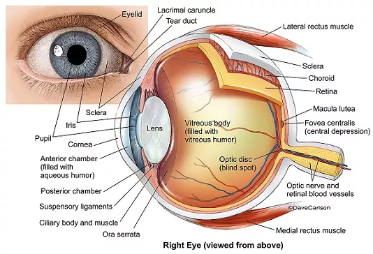
To help with eye anatomy, it is useful to differentiate the eye into 3 parts:
– Front compartment
– Back compartment
– Surrounding supporting tissues
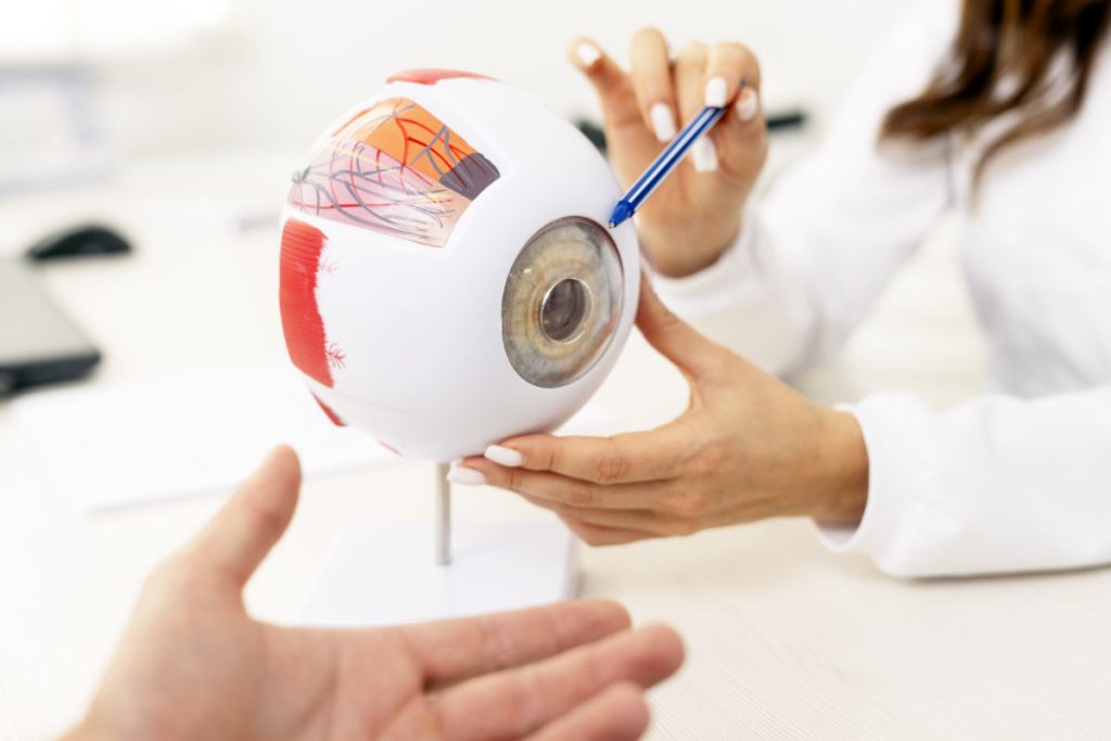
Model eyes are very useful to help with identifying different parts of the eye anatomy. Below are a selection of model eyes (and of the brain too, if you are interested) that are available for purchase through Amazon, ranging from fairly basic to very detailed:
What structures are at the front of the eye?
The front of the eye consists of the following tissues: conjunctiva, sclera, cornea, iris, ciliary body and crystalline lens. The main job is to allow light to enter the eye and to focus this light onto the retina.
The conjunctiva is the thin layer that lines the front of the sclera and also the inner surfaces of the eyelids. It contains glands that secrete lubricating fluids as well as lymphoid tissue. The conjunctiva is important in keeping the eye moist and in protecting the eye from infection. Dry eye syndrome occurs when there are problems with the conjunctiva.
The sclera is the tough outer wall of the eye. It is the white part of the eye. The sclera maintains the structure of the eye and provides support for all the other eye tissues. The eye movement muscles are attached to the sclera – anchor points on the sclera allow the eyeball to move when the eye movement muscles contract.
The cornea is the clear, front window of the eye. It is transparent; this allows light to enter the eyeball. As light passes through the cornea, it becomes refracted. Together with the lens, the cornea is responsible for focusing light onto the retina.
The space in between the cornea and lens is called the anterior chamber, which is filled by aqueous humor. The aqueous humor provides nutrients and oxygen to the cornea.
The iris is the colored part of the eye. It is the membrane that sits between the cornea and the lens. The iris color depends on the amount of pigment in the iris. Those with dark brown irides have a lot of iris pigment.
The pupil is the round opening in the center of the iris. The iris is responsible for controlling the amount of light that enters the eye through the pupil. When there is too much light, the iris muscles constrict the pupil to reduce the amount of light entering the eye. When it is dark, the iris muscles dilate the pupil to allow as much light to enter the eye as possible.
The crystalline lens is located just behind the iris. It is held in place in the eye by zonules (also known as suspensory ligaments) that attach it to the ciliary body. Together with the cornea, it is responsible for focusing light onto the retina. Changing its thickness allows you to change focus from far to near and vice versa (i.e. accommodate).
As you grow older, this ability to change the focusing distance diminishes and you end up needing to wear reading glasses – this is called presbyopia. Age also causes the lens to become progressively more cloudy, resulting in cataract formation.
The ciliary body is positioned just behind the iris. Its two main functions are to produce aqueous humor and to change the thickness of the lens by stretching or relaxing the zonules that are attached to it.
And the structures at the back of the eye?
The back compartment consists of the following: vitreous humor, retina, choroid and optic nerve.The main job is to allow light entering the eye to reach the retina, and to send the light signals to the brain for analysis.
The vitreous humor is the jelly-like substance that fills the large back cavity between the lens and the retina. It is transparent and allows light to be focused onto the retina. The vitreous humor also helps the eye to maintain its shape and turgidity. Vitreous loss into the anterior chamber is one of the complications that can occur during cataract surgery.
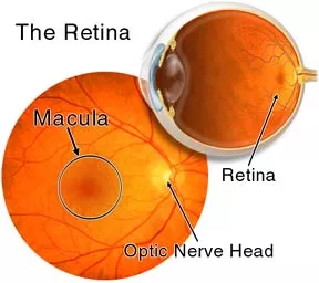
The retina is the light-sensitive innermost lining of the eyeball. It is probably easiest to think of the retina as wallpaper in a room.
The retina consists of the retinal pigment epithelial layer and neurosensory retina. The retinal pigment epithelium helps to maintain and support the neurosensory retina. The neurosensory retina detects, absorbs and processes the light that enters the eye. Retinal photoreceptor cells in the neurosensory retina collect light signals and convert them to bio-electrical signals, which are then sent to the optic nerve.
There are two types of photoreceptor cells: rods and cones.
Rod photoreceptors are located in the peripheral retina, and are responsible for night vision and detection of motion.
Cone photoreceptors are mostly concentrated at the macula (center of the retina) and are important for fine detailed visual acuity and color vision. Any damage to the macula, such as in macular degeneration, will cause blurring of the central vision.
The optic nerve is the cable connecting the eye to the brain. It is composed of well over one million nerve fibers. It transmits bio-electrical information from the neurosensory retina to the brain, so that the brain can interpret what it is that you are seeing. Your vision can be affected by damage to the optic nerve, such as in glaucoma and optic neuritis.
The choroid is the layer between the retina (wallpaper) and sclera (wall). It is full of blood vessels and is responsible for supplying nutrients to the retina. The dark melanin pigment in the choroid absorbs any excess light in the eye.
What about supporting structures of the eye?
We shouldn’t forget the important supporting structures of the eye, which nclude the eyelids, eye movement muscles and lacrimal drainage system.
The eyelids protect the eyes from any external trauma. Blinking the eyelids maintains the tear film, which keeps the eye moist. The upper and lower eyelids have a tough fibrous plate called the tarsal plate, which provides support and rigidity to the lid. These plates contain meibomian glands, which produce oil that helps to stabilize the tear film. Dysfunction of the meibomian glands can result in inflammation of the eyelids and blepharitis.
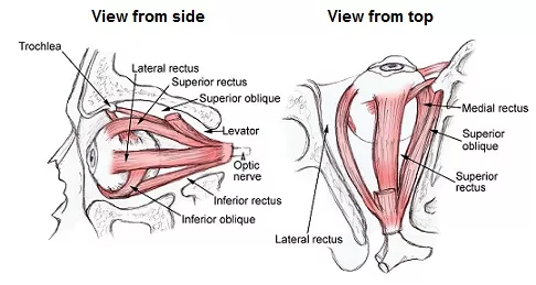
The extraocular (eye movement) muscles move the eye. There are six muscles that connect the sclera to the eye socket. The six muscles are the superior rectus, inferior rectus, lateral rectus, medial rectus, superior oblique and inferior oblique muscles. These muscles work together so that both eyes will view an object at the same time.
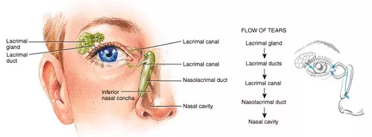
The lacrimal system produces, distributes and drains the tears that keep the surface of the eye moist. The lacrimal gland is located under the upper eyelid and its job is to produces tears.
When you blink, the tears are distributed throughout the eye surface. The tears are then drained through the punctum, lacrimal canaliculi, nasolacrimal sac and nasolacrimal duct to eventually enter the nose.
Where can I learn more?
For all you budding optometrists and ophthalmologists out there, the best eye anatomy book in my opinion is Clinical Anatomy of the Eye by Richard Snell and Michael Lemp. The book is very clearly and concisely written, and has many diagrams and images that illustrate the anatomy well.
I should know because this was the book that got me through my ophthalmology postgraduate specialist training anatomy exam! Very highly recommended if you want to learn more about the anatomy of the eye.
