The eyes are amazing organs that have been designed to provide a variety of visual functions. Most of the time, you will just accept what you see and not think about how much you perceive visually. When your eyes work well, all the components of visual function merge together effortlessly to give you your sight.
Take a few moments to think about this. You are able to read fine print at close distances. Yet you can also see distant objects relatively clearly. But the objects that you see are not just black and white monochrome. They have color!
And you don’t just see objects that are directly ahead of you – you can also perceive things that are around you. Wait there’s more! You can perceive movement! Things don’t just appear stationary. Finally, ever notice that you can see well in bright conditions and also in dark conditions?
What are the main visual function tests?
The visual processing that occurs from the eye to the brain is very complex and is still not completely understood. But there are ways for your vision and visual function to be tested and measured. The commonest visual function tests that are performed at clinics and hospitals are: visual acuity, visual field, color vision and contrast sensitivity.
Visual acuity is essentially the clarity and sharpness with which you see things, i.e. how well you can see. It is a measure of how well your eye and your brain can distinguish spatial resolution. Your entire visual pathway from the eye to the brain, needs to be in good working order for you to maintain good visual acuity.
Test your visual acuity with your free Snellen chart
Color vision is the ability to appreciate differences in color, as opposed to just black, white and shades of gray. The color of an object depends on the wavelength of the light that the object reflects or emit. When the light enters the eye, it is detected by the cone photoreceptor cells in the retina. Light of different wavelengths will stimulate different cone photoreceptors. The interaction between the cone photoreceptors and the subsequent processing in the brain provides the perception of color and gives you color vision.
The 2 main ways of assessing the ability to appreciate color are with the Ishihara and Farnsworth-Munsell tests.
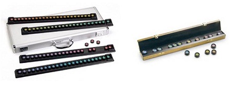
The Farnsworth-Munsell color tests: the 100 Hue Test (left) and the D-15 test (right)
Your visual field, in practical clinical terms, is essentially the area of space that can you can see at the same time. In other words, it refers to your field of vision or how far out peripherally you are able to see without moving your eyes or head. Generally speaking, if light that is reflected or emitted from an object in your surroundings falls onto your retina, then that object will be visible in your visual field.
The 2 main ways of evaluating your visual fields are with static perimetry and kinetic perimetry. In static perimetry, points of light (at fixed locations) are flashed at different brightness levels until you see them. In kinetic perimetry, points of light (of different sizes and brightness) are moved inwards slowly one by one until you see them.
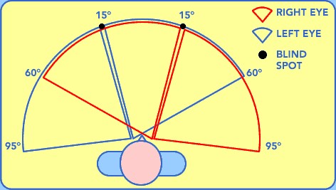
The diagram above depicts the normal horizontal field of vision, including the location of the blind spots for both eyes. In a normal person, the field of vision should span a total width of 190 degrees.
Contrast sensitivity refers to the ability to distinguish an object from other objects and the background, even when it is not clearly outlined or prominent. Maximum contrast is obtained when comparing between black and white. However, when comparing between shades of gray, your ability to discern outlines becomes reduced.
When you have poor contrast sensitivity, you will be unable to see small contrast differences between adjacent objects and surfaces. For example, you will find it difficult to see facial expressions. You may find navigating steps difficult because you are unable to recognize the edges of the steps. Interestingly, you can have good visual acuity but yet have poor contrast sensitivity.
Eye diseases that can cause reduced contrast sensitivity include cataract, glaucoma, macular degeneration, diabetic retinopathy and optic neuritis. Contrast sensitivity can be measured with the Pelli-Robson chart or Vistech contrast sensitivity test.
Stereopsis is the ability of both eyes to see the same object as one image and to create a perception of depth. It is a measure of binocular visual function, i.e. how well both eyes work together. Stereopsis allows you to judge distances and to see where objects are in relation to you and to each other.
Stereoscopic depth perception is measured in seconds of arc. In general, you are considered to have gross stereoscopic vision at 3,600 seconds of arc. The smaller the number (some people can achieve stereovision better than 20 seconds of arc), the better your stereopsis. Stereopsis is can be assessed with contour stereotests (such as the Titmus Fly stereotest) and random-dot stereograms (such as the Frisby, Lang, TNO and randot stereotests).
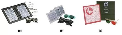
a) Randot stereotest; (b) Random-dot E stereotest; (c) TNO stereotest
