What is stereopsis?
Stereopsis, also known as stereoscopic depth perception, is the ability of both eyes to see the same object as one image and to create a perception of depth. It is a measure of binocular visual function, i.e. how well both eyes work together.
While not as commonly tested for in adults compared to the uniocular visual function tests (such as visual acuity, color vision and visual fields), it is nevertheless a very important part of how you see in your day-to-day living. Having stereopsis allows you to judge distances and to see where objects are in relation to you and to each other.
Is it really that important? Well, yes. You need stereoscopic depth perception to be able to perform these tasks well:
– Driving and parking your car
– Pouring a cup of tea
– Threading a needle, sewing and knitting
– Climbing up and down stairs
– Reaching out to touch or hold something
– Suturing and performing surgery (if you want to become a surgeon)

Are these examples of purely bad parking by bad drivers or examples of poor stereopsis?
Most of the time, poor or absent stereopsis is due to childhood amblyopia. However, stereopsis can also be affected later on in life by conditions that reduce your ability to see clearly, for example cataract, age-related macular degeneration and presbyopia. Finally, bear in mind too that disorders of the brain (particularly if the visual processing pathways are involved, such as in a stroke or head injury) will have an effect on your stereopsis.
How is stereopsis perceived?
Before going into greater detail about stereoscopig depth perception, it is important to understand a little bit more about binocular vision and how your eyes work together.
Binocular vision refers to the ability of both eyes to work together to see the same object simultaneously. But that’s not all there is to it. Both your eyes do not see things the same way as each other, i.e. the image obtained in one eye differs from the other. The only way that the images from both eyes can be the same is if both your eyes are located in the same position. Which of course, is impossible unless you are a cyclops (and even then, a cyclops only has one eye…).
If your brain interprets the images that both eyes see as separate images, you will develop double vision (or diplopia). To get round this problem, one of two things can happen: suppression or fusion.
Suppression occurs when the brain actively suppresses or ignores the image from one eye. Typically, the images from the non-dominant eye are suppressed. This way, only images from the dominant eye are processed by the brain, and double vision is avoided. In children under 8 years of age, this suppression can lead to reduced vision from amblyopia because the visual pathways between the suppressed eye and brain are not continuously being developed. Amblyopia treatment therefore requires patching of the good eye in order to stimulate visual pathways of the suppressed non-dominant eye.
Fusion occurs when the brain is able to bring together the two disparate images from both eyes and interpret them as one single image. This is what usually happens for normal eyes and normal visual processing systems in the brain. You need to have fusion first before you can have depth perception.
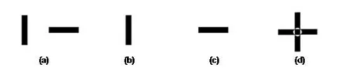
a) Testing for fusion: a vertical bar is presented to the left eye and a horizontal bar to the right eye. What is seen with both eyes open?
(b) Only the vertical bar is seen. This indicates that the image from the right eye has been suppressed, i.e. suppression of the right eye has occurred.
(c) Only the horizontal bar is seen. This indicates that the image from the left eye has been suppressed, i.e. suppression of the left eye has occurred.
(d) A ‘plus’ sign is seen. Images from both eyes have been fused to form a single image.
As your eyes are located closely adjacent to each other, they are able to see the same objects simultaneously but at a slightly different angle to each other. Thus, there are slight differences between the images that have been captured by each eye.
During the fusion process, the 2 disparate images are combined into one. Similarities between the 2 images are matched together, and more importantly, the slight disparities are also added in. It is this that gives you your ability to perceive depth and appreciate 3D.
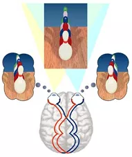
Note: Fusion can only occur if the disparities in the images captured by both eyes are small. If the difference between the images is too great, then either double vision or suppression occurs.
How is stereopsis measured?
Stereoscopic depth perception is measured in seconds of arc. In general, you are considered to have gross stereoscopic vision at 3,600 seconds of arc. The smaller the number (some people can achieve stereovision better than 20 seconds of arc), the better your stereopsis.
Stereovision tests are primarily used in children, as a vision screening tool for amblyopia and binocular vision defects, and also as a way of monitoring the progress of amblyopia treatment. There are 2 groups of clinical tests (also called stereotests) that are used to measure stereopsis: contour stereotests and random-dot stereograms.
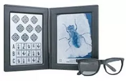
Contour stereotests use two horizontally disparate images to evaluate stereopsis. An example is the Titmus Fly stereotest (left). After wearing polarizing spectacles (so that each eye sees a different image), you will need to determine which of the images have depth.
Random-dot stereograms were designed to eliminate monocular cues from depth perception testing, to improve the accuracy of stereovision testing. Examples include the Randot stereotest, Random-dot E stereotest, TNO stereotest, Frisby stereotest and the Lang stereotest. All these tests require you to correctly identify the target or image that has stereoscopic depth at a set distance (usually 40 centimeters) from your eyes.
The Randot and Random-dot E stereotests utilize polarizing vectographs and so require the wearing of polarizing glasses before testing can be performed (similar to the Titmus fly stereotest). On the other hand, the TNO stereotest uses the principle of red-green dissociation, and thus red-green glasses (instead of polarizing glasses) are required.
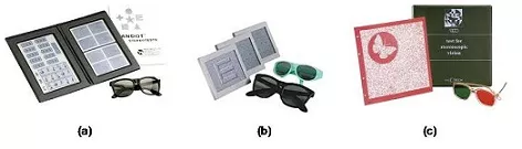
(a) Randot stereotest; (b) Random-dot E stereotest; (c) TNO stereotest
The 2 random dot stereograms that do not require glasses are the Frisby and Lang stereotests. Both use different principles to avoid the need for glasses during testing. The Frisby stereotest uses a series of squares containing geometric shapes painted on perspex of different thicknesses. The Lang stereotest uses a combination of random dots and cylinder gratings.
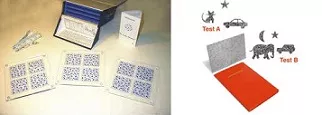
Left: Frisby stereotest. Right: Lang stereotest. The advantages of not requiring glasses include easier testing in uncooperative children who are adamant about not wearing glasses and unimpeded observation of the child’s eye movement behaviour by the examiner.
